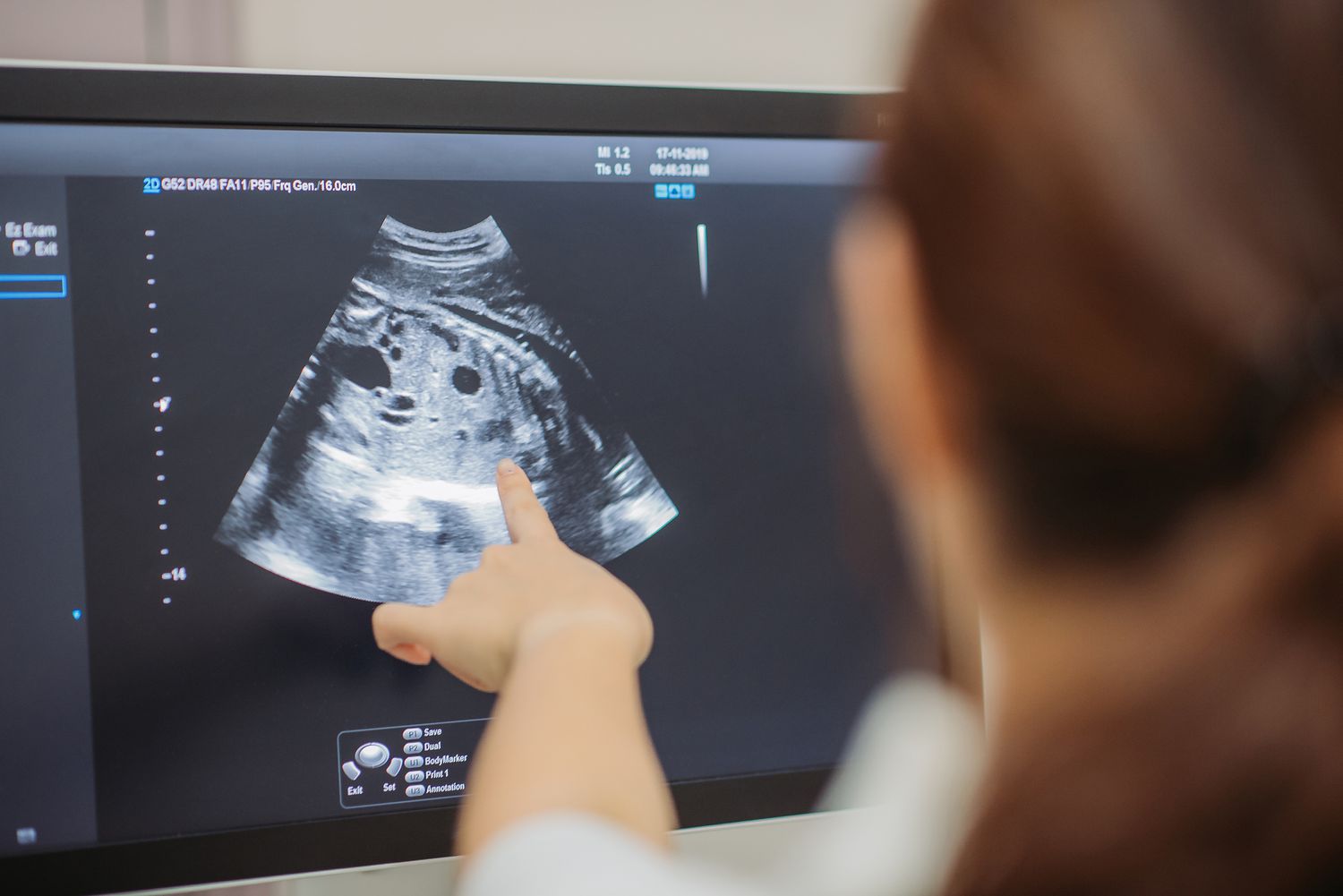
Pregnancy is a time of anticipation and excitement, filled with moments of wonder and curiosity about the life growing inside the womb. Among the many prenatal tests and examinations, fetal echocardiography stands out as a remarkable tool, allowing parents and healthcare professionals to gain insights into the developing heart of the unborn child. In this blog post, we’ll explore what fetal echocardiography is, why it’s essential, and what to expect during this examination.
What Is Fetal Echocardiography?
Fetal echocardiography, often simply called a “fetal echo,” is a specialized ultrasound examination that focuses on the development and health of the fetal heart. It provides a detailed view of the baby’s heart, including its structure and function. This non-invasive procedure is typically performed between the 18th and 24th weeks of pregnancy, offering crucial information about the heart’s condition before birth.
Why Is Fetal Echocardiography Important?
- Early Detection of Heart Abnormalities: Fetal echocardiography can identify congenital heart defects (CHDs) early in pregnancy. CHDs are among the most common birth defects and can vary in complexity, from simple to complex. Early detection allows for appropriate medical planning and intervention.
- Informed Decision-Making: A diagnosis of a CHD during pregnancy allows parents and healthcare providers to make informed decisions about the baby’s care, delivery, and potential post-birth treatments.
- Specialized Care: If a CHD is detected, the medical team can prepare for the baby’s arrival, ensuring that specialized care and interventions are available immediately after birth.
- Reduced Stress and Anxiety: Fetal echocardiography can provide reassurance when no heart abnormalities are found, reducing stress and anxiety for expectant parents.
What to Expect During Fetal Echocardiography
During a fetal echo, the procedure is generally similar to a regular prenatal ultrasound. Here’s what you can expect:
- Gel Application: A water-based gel will be applied to the mother’s abdomen to create better contact between the transducer and the skin.
- Transducer Placement: A specialized transducer will be moved over the abdomen to capture images of the fetal heart.
- Image Analysis: The sonographer or specialist will analyze the images in real-time, focusing on the heart’s structure, function, and blood flow.
- Duration: The procedure typically takes about 30 minutes, but it may vary based on the complexity of the examination.
- Results: The results are usually discussed immediately after the procedure, allowing parents to ask questions and understand the findings.
Conclusion
Fetal echocardiography is a remarkable and essential diagnostic tool that offers parents a glimpse into their baby’s heart health during pregnancy. Whether the examination provides reassurance or leads to early intervention, it plays a crucial role in ensuring the best possible outcome for babies with congenital heart defects. As an expectant parent, being informed about the process and its significance empowers you to make the best decisions for your child’s future. Remember, the ability to see your baby’s heart is more than just an image on a screen; it’s a window into a future filled with care and hope.


